Top Guidelines Of Doppler Ultrasound Exam Of An Arm Or Leg Information - Mount ...
( Some issues may not be selected up by this scan.) Sometimes, it is extremely difficult to examine child through the abdominal area due to bowel gas or the thickness of tissue under the skin. Increased fat frequently makes it more difficult to see the infant plainly. An internal scan can be the very best alternative to get a better take a look at baby.
A firm wedge is then put under the lady's bottom to raise the hips. The transducer is a long tubular structure with a deal with. It is covered with a condom and sterile gel is put on the pointer so that it can be positioned into the vaginal area quickly. If you consent to this, the transducer is placed into the vagina, which offers a clearer view of baby.
It is necessary to let the sonographer understand if you feel any pain. This treatment is unpleasant but must not hurt Trans-vaginal scan are typically done in early pregnancies for dating scans. Ultrasound can identify some kinds of physical abnormality. Examples of physical birth defects that may be found at 19 - 20 weeks are most cases of spina bifida, some severe heart defects, some kidney problems, lack of part of a limb and some cases of cleft palate.
What Does Ultrasound Scan - Tests And Scans Do?
This unpredictability or 'not knowing' may trigger anxiety. Your medical professional or midwife can provide more information and support. Ultrasound can typically show the child's gender, but it is not constantly 100% ensured. You may choose whether or not you want to be told. The results will be sent out to the medical professional who referred you to have the scan.
Other tests may be required to get more details. These tests may consist of a more scan at a later date or a test to analyze the baby's chromosomes ' Screening tests for Down syndrome'. diagnostic ultrasound. Having a regular result on an ultrasound scan does not ensure that your baby will not have an abnormality or chromosomal abnormally.
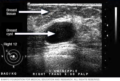
Ultrasound is an imaging test that sends out high-frequency acoustic waves through your breast and transforms them into images on a viewing screen. The ultrasound service technician places a sound-emitting probe on the breast to conduct the test. There is no radiation involved. Ultrasound is not utilized on its own as a screening test for breast cancer.
How Chest Ultrasound can Save You Time, Stress, and Money.
If an irregularity is seen on mammography or felt by physical examination, ultrasound is the very best method to discover out if the abnormality is solid (such as a benign fibroadenoma or cancer) or fluid-filled (such as a benign cyst). It can not identify whether a strong lump is malignant, nor can it detect calcifications.
Mammograms can be challenging to translate in young ladies since their breasts tend to be thick and loaded with milk glands. (Older females's breasts tend to be more fatty and are easier to evaluate.) In mammograms, this glandular tissue looks dense and white much like a cancerous growth. Some doctors say that locating an abnormality in the middle of thick gland tissue can be like finding a polar bear in a snowstorm.
Doctors likewise can utilize ultrasound to assist biopsy needles specifically to suspicious areas in the breast. Was this article helpful? Yes/ No Last customized on December 14, 2020 at 3:45 PM.
Ultrasound Imaging Can Be Fun For Anyone
 Hypertension is the most often dealt with disease this page in internal medicine - diagnostic ultrasound surrey. More than 1 billion people worldwide struggle with hypertension. High blood pressure results in cardiovascular end-organ damage increasing morbidity and mortality and is related with high expenses to society, making this illness an important public health challenge. Sonography is a vital diagnostic tool in the evaluation of a hypertensive patient.
There are a number of ultrasound assessments that may be required in hypertension. Stomach ultrasound is recommended by several guidelines for the basic diagnostic workup in every newly diagnosed hypertensive client. Doppler sonography of the kidney arteries is affordable just in a subset of hypertensives that are at increased threat of kidney artery stenosis.
Ultrasound of the carotid arteries is often used to detect and evaluate in the case of hypertension-induced vascular end organ damage. The evaluation of the intima-media density permits the detection of early phases of atherosclerotic wall changes. Prior to any structural vascular damage that may be pictured by ultrasound methods, hypertension results in practical modifications of the endothelium, called endothelial dysfunction.
The Ultrasound Imaging Statements
This can be detected by sonography determining the size changes of the brachial artery in reaction to predefined endothelial stimuli. Flow-mediated dilation in action to hyperemia is considered as the gold-standard in the non-invasive assessment of endothelial dysfunction. To date, it is rather used clinically than in day-to-day scientific practice.
Using abdominal ultrasound in the examination of high blood pressure is twofold. In the detection of a secondary forms of hypertension. In the assessment of subclinical organ damage caused by high blood pressure. In the existing European Society of Cardiology/European Society of Hypertension (ESC/ESH) guidelines for hypertension using abdominal ultrasound is recommended as a part of the evaluation of hypertensive people.
The primary interest is the morphology of the kidneys, the adrenal glands and of the aorta. Due to their retroperitoneal position, kidneys are totally and quickly noticeable. A 3. 5-5 MHz probe is this content generally used to scan the kidney. The evaluation from dorsolateral permits the evasion of the digestive loops and hence enables a non-overlapping imaging in the supine position.
Some Known Factual Statements About What Is A Medical Ultrasound?
Kidney ultrasound has now practically entirely changed intravenous urography in the anatomical expedition of the kidney. While the latter needs the injection of potentially nephrotoxic contrast medium, ultrasound is non-invasive and provides the necessary anatomic information about kidney size and shape, cortical thickness, urinary tract blockage and kidney masses [1].
The finding of bilateral upper stomach masses at physical assessment follows polycystic kidney disease and must require an abdominal ultrasound assessment. Intense parenchymal inflammatory processes like crescentic glomerulonephritis or intense interstitial nephritis in some cases inclines individuals to quantifiable organ swelling. The cortical and medullary pyramids have in this case an anechoic profile.